The Liver and Gallbladder: Biliary Physiology, Fat Emulsification, and Metabolic Detoxification
An exhaustive clinical exploration of liver and gallbladder physiology. Discover bile acid synthesis, lipid emulsification, enterohepatic circulation, hepatic lobule architecture, and phase I/II detoxification.
disclosure This article contains affiliate links. Purchases may earn us a commission at no extra cost to you. We only recommend products we've reviewed.
The Liver and Gallbladder: Biliary Physiology, Fat Emulsification, and Metabolic Detoxification
While the stomach functions as the body's primary acid-driven biochemical reactor, the transition of food into the small intestine requires an immediate and radical shift in physiological strategy. The highly acidic, nutrient-dense chyme exiting the pyloric sphincter cannot be processed or absorbed in its raw state. To prevent chemical burns to the intestinal epithelium and to unlock the energy stored within dietary fats, the human body relies on the largest internal organ and its dedicated storage vesicle: the liver and the gallbladder.
Together, the liver (hepar) and gallbladder form an indispensable hepatobiliary axis. Working in synchronized harmony, these organs synthesize, concentrate, store, and secrete a complex biological fluid known as bile. Far beyond its role as a simple digestive juice, bile serves as a critical dual-function secretion: it is an essential detergent that emulsifies dietary lipids for absorption and a major excretory pathway for metabolic waste products, neutralized xenobiotics, and degraded hemoglobin.
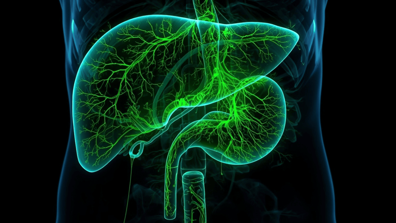 Detailed anatomical illustration of the hepatobiliary tree, illustrating bile flow from intrahepatic ducts to the gallbladder and common bile duct.
Understanding the physiological mastery of the liver and gallbladder requires diving deep into micro-anatomical architecture, enzymatic conversion pathways, and complex neuro-hormonal feedback loops. As established in our Complete Clinical Guide to the Human Digestive System, the hepatobiliary system represents the central metabolic clearance house and lipid-processing powerhouse of human physiology, bridging the gap between raw gastric digestion in the The Stomach: Clinical Physiology, Gastric Acid Dynamics, and Protein Digestion and systemic nutrient assimilation.
1. Functional Micro-Anatomy: The Hepatic Lobule, Portal Triads, and Biliary Tree Architecture
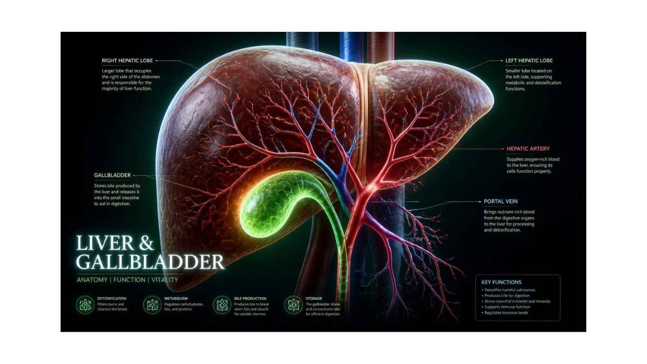
To comprehend how the liver processes roughly 25% of total cardiac output every single minute, one must examine its functional histological unit: the hepatic lobule. Under microscopic examination, the liver architecture is organized into hundreds of thousands of hexagonal lobules, each structured around a central vein that ultimately drains into the hepatic veins and inferior vena cava.
At each of the six corners of a hexagonal hepatic lobule lies a specialized functional structure known as the Portal Triad. The portal triad contains three distinct micro-vessels encased in connective tissue:
- A Branch of the Portal Vein: Delivers nutrient-rich, deoxygenated blood directly from the stomach, intestines, spleen, and pancreas (accounting for roughly 75% of total hepatic blood flow).
- A Branch of the Hepatic Artery: Delivers oxygenated blood directly from the systemic circulation via the celiac trunk (accounting for the remaining 25% of hepatic blood flow).
- A Bile Ductule: A lined epithelial conduit that collects newly synthesized bile flowing in the opposite direction from surrounding hepatocytes.
Blood from both the portal vein branch and hepatic artery branch enters specialized, low-pressure fenestrated capillaries called hepatic sinusoids. These sinusoids radiate inward toward the central vein, lined by specialized endothelial cells and filled with Kupffer cells—resident tissue macrophages that filter out senescent red blood cells, immune complexes, and blood-borne pathogens coming from the intestinal tract.
Running parallel to these blood-filled sinusoids, but isolated by impermeable tight junctions, are microscopic channels called bile canaliculi. Hepatocytes lie arranged in double-cell plates between the sinusoids and canaliculi. This precise spatial arrangement creates a counter-current flow system: blood flows inward from the portal triads toward the central vein, while newly synthesized bile flows outward through the canaliculi toward the bile ductules at the portal triads.
As bile exits the microscopic canaliculi, it enters larger interlobular bile ducts, which gradually coalesce into the left and right hepatic ducts. These two major conduits exit the liver porta hepatis and unite to form the Common Hepatic Duct. From here, bile can either flow straight down the Common Bile Duct into the duodenum or divert via the Cystic Duct into the gallbladder for concentration and storage.
2. Bile Acid Biochemistry: Cholic Acid Synthesis, Conjugation, and Micellar Dynamics
Bile is a complex aqueous solution composed of water, electrolytes, bile salts, phospholipids (primarily phosphatidylcholine/lecithin), cholesterol, bilirubin, and trace heavy metals. The most critical functional components of bile regarding lipid digestion are bile salts.
The synthesis of bile acids represents the primary pathway for cholesterol clearance in the human body. Hepatocytes synthesize primary bile acids—specifically cholic acid and chenodeoxycholic acid—directly from cholesterol through a complex series of enzymatic reactions initiated by the rate-limiting enzyme Cholesterol 7α-hydroxylase (CYP7A1).
Because native cholesterol and un-conjugated bile acids have poor water solubility at physiological pH, hepatocytes perform a crucial enzymatic conjugation step before secreting them into bile canaliculi. Primary bile acids are conjugated with either the amino acid glycine or taurine at a ratio of roughly 3:1. This biochemical conjugation significantly lowers the molecule's dissociation constant ($pK_a$), converting insoluble bile acids into fully ionized bile salts (glycocholic acid, taurocholic acid, glycochenodeoxycholic acid, and taurochenodeoxycholic acid).
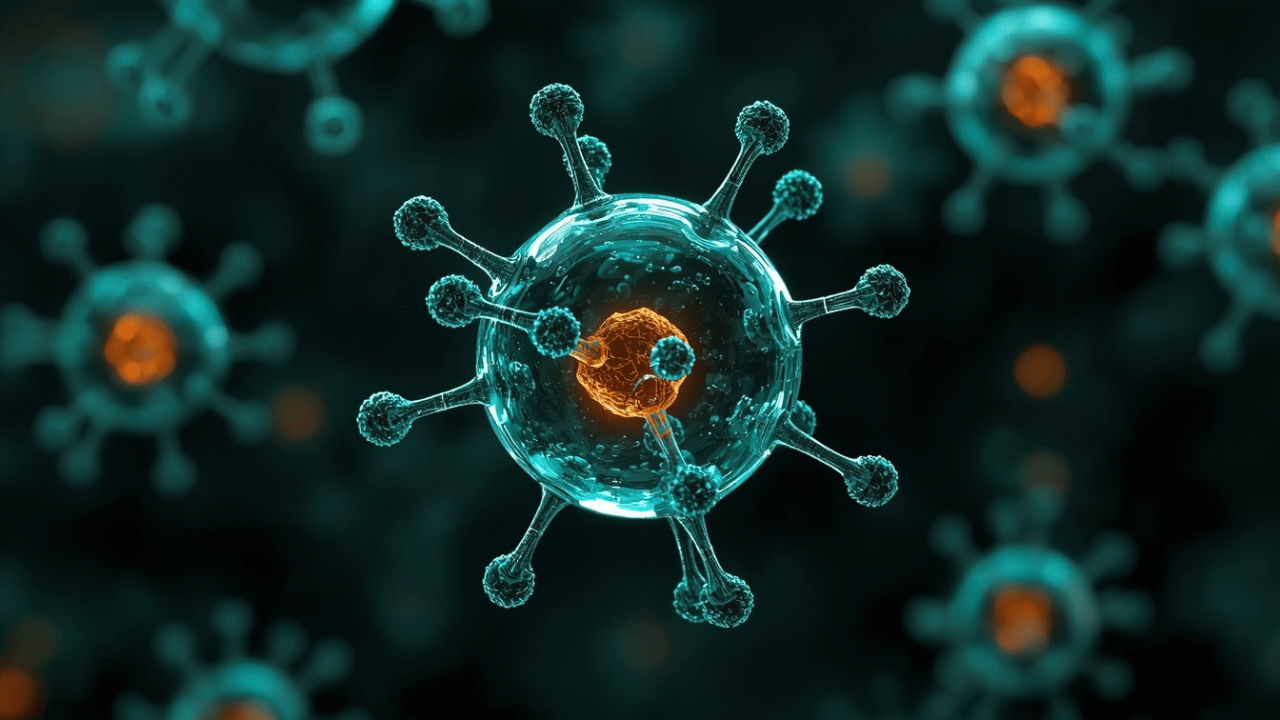 3D molecular illustration of an amphipathic bile salt molecule showing its hydrophilic and hydrophobic faces assembling into a spherical lipid micelle. *
The defining physiological characteristic of a bile salt is its amphipathic structure. Unlike typical molecules that have distinct polar ends, a bile salt molecule possesses a distinct hydrophobic (fat-soluble) steroid ring face and a hydrophilic (water-soluble) hydroxyl and amino acid face.
This dual nature allows bile salts to function as powerful bio-detergents. When introduced into an aqueous environment containing large dietary fat globules, the hydrophobic faces of bile salts align with the non-polar triglycerides and cholesterol, while their hydrophilic faces interact with the surrounding water molecules.
As mechanical churning in the stomach and small intestine breaks large fat drops into smaller droplets, bile salts rapidly coat these newly created surfaces. This coating prevents small lipid droplets from coalescing back into large, insoluble fat masses—a mechanical process known as emulsification.
By breaking large fat drops down into millions of microscopic emulsification droplets, bile salts expand the available surface area of dietary lipids by more than 1,000-fold. This drastic increase in surface area enables the water-soluble enzyme Pancreatic Lipase (working alongside its co-factor, colipase) to rapidly dock onto lipid surfaces and hydrolyze triglycerides into free fatty acids and 2-monoacylglycerols.
As lipolysis progresses, bile salts, phospholipids, monoacylglycerols, free fatty acids, cholesterol, and fat-soluble vitamins ($A$, $D$, $E$, $K$) self-assemble into tiny aggregate structures called mixed micelles (measuring 3 to 10 nanometers in diameter). These mixed micelles act as molecular transport vehicles, diffusing across the unstirred water layer of the intestinal lumen to deliver hydrophobic lipid breakdown products directly to the brush border membrane of enterocytes for passive and carrier-mediated absorption.
3. The Gallbladder: Concentrating Dynamics, Cholecystokinin (CCK) Neuro-Hormonal Cascade, and Motility
The gallbladder is a pear-shaped, muscular sac nestled in a shallow fossa on the inferior surface of the liver's right lobe. Anatomically divided into a fundus, body, infundibulum, and neck, its primary physiological duty is not synthesis, but the storage, concentration, and timed delivery of hepatic bile.
The liver synthesizes bile continuously at a rate of 500 to 1,000 milliliters per day. However, during fasting periods between meals, the Sphincter of Oddi—a smooth muscle valve surrounding the Hepatopancreatic Ampulla of Vater where the common bile duct enters the duodenum—remains tightly closed. This closure creates a backpressure gradient within the biliary tree, forcing newly formed hepatic bile to flow backward through the cystic duct into the gallbladder.
Once inside the gallbladder lumen, bile undergoes a dramatic physiological concentration process:
- Epithelial Transport Mechanics: The specialized columnar epithelium lining the gallbladder wall actively transports sodium ($Na^+$) and chloride ($Cl^-$) ions out of the bile lumen and into the basolateral intercellular spaces utilizing $Na^+/K^+$ ATPase pumps and $Na^+/H^+$ exchangers.
- Isosmotic Water Reabsorption: As electrolytes are continuously pumped out of the lumen, water passively follows down its osmotic gradient through transcellular aquaporin channels and paracellular pathways, returning directly to the surrounding capillary blood supply.
- Bile Concentration: Over a fasting period of several hours, this active fluid extraction concentrates the organic solid components of bile (bile salts, bilirubin, cholesterol, and lecithin) by 5 to 20 times, turning dilute hepatic bile into a viscous, concentrated dark-green storage fluid.
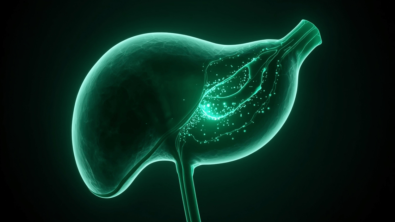 Clinical 3D visual of gallbladder smooth muscle contraction releasing concentrated bile through the cystic duct upon cholecystokinin (CCK) stimulation.
The release of concentrated gallbladder bile into the small intestine is governed by a precise neuro-hormonal reflex cascade triggered by meal ingestion:
- Cephalic and Gastric Phases: Vagal nerve stimulation (acetylcholine) during the sight, smell, and stomach distension of a meal causes mild gallbladder contraction and partial relaxation of the Sphincter of Oddi, preparing the biliary tree for action.
- Intestinal Phase (The Master Trigger): As acidic chyme containing partially digested proteins and long-chain fatty acids enters the duodenum, specialized neuroendocrine cells in the duodenal mucosa called I-cells detect these lipid molecules and rapidly secrete the peptide hormone Cholecystokinin (CCK) into the bloodstream.
- Targeted CCK Response: Circulating CCK binds to specific $CCK_1$ receptors on the smooth muscle wall of the gallbladder, inducing powerful, rhythmic contractions. Simultaneously, CCK binds to receptors in the sphincter of Oddi, signaling the smooth muscle ring to completely relax.
- Bile Ejection: This coordinated contraction-relaxation pathway generates a strong pressure gradient, driving a concentrated surge of bile out of the gallbladder, down the common bile duct, and through the open Ampulla of Vater directly into the duodenal lumen to mix with incoming dietary fats.
4. Enterohepatic Circulation: Biliary Recycling and Microbial Biotransformation
The human body contains a total bile acid pool of approximately 2 to 4 grams. However, efficient lipid digestion during a typical day requires between 12 and 30 grams of bile salts. To meet this massive biochemical demand without requiring the liver to constantly synthesize vast quantities of new cholesterol derivative molecules, the gastrointestinal tract relies on an extremely efficient recycling system known as Enterohepatic Circulation.
Over 95% of the bile salts secreted into the duodenum are successfully reabsorbed and returned to the liver, while less than 5% are lost in the feces daily (a loss that precisely matches the rate of new hepatic bile acid synthesis from cholesterol).
A dynamic visual diagram mapping this precise enterohepatic loop would reveal the following continuous pathway:
- Hepatocytes synthesize bile and secrete it into bile canaliculi.
- Bile flows through the biliary tree to the gallbladder for storage and concentration.
- CCK triggers gallbladder ejection into the duodenal lumen for fat emulsification.
- Bile salts travel through the full length of the small intestine.
- Upon reaching the terminal ileum, specific transporters reabsorb 95% of the bile pool.
- The reabsorbed bile salts enter the portal venous circulation and travel back to the liver to be cleared and re-secreted.
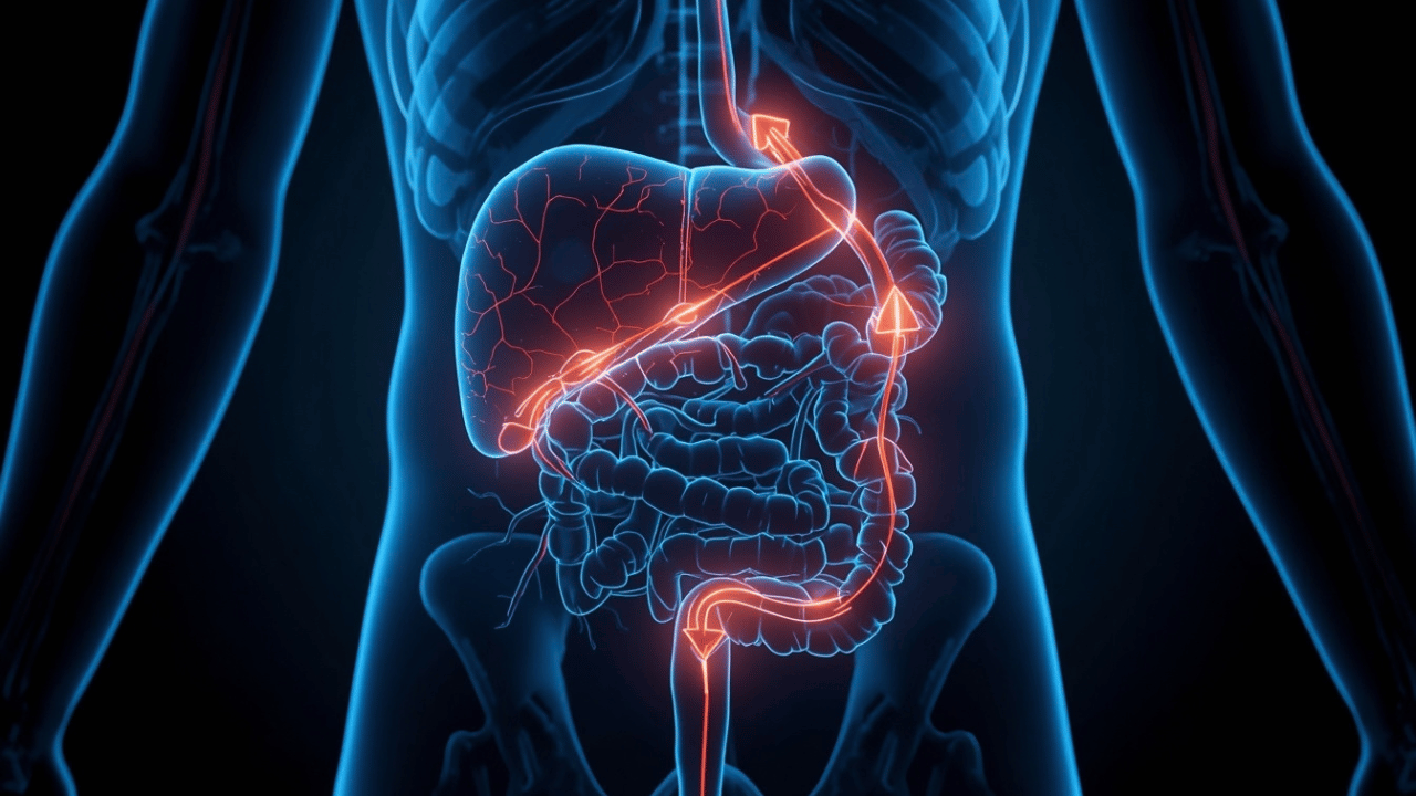
Clinical 3D anatomical map illustrating enterohepatic circulation: bile transport from liver to ileum and portal venous return.
The recycling of bile salts takes place through active transport across the mucosal epithelium. Inside the ileal enterocyte, bile salts are exported directly into the portal venous circulation.returning bile salts travel through the portal vein back to the liver. On reaching the hepatic sinusoids, hepatocytes extract bile salts from sinusoidal blood with near 90% first-pass efficiency, immediately re-secreting them back into bile canaliculi.
Microbial Biotransformation in the Colon
The 5% of bile salts that escape reabsorption pass into the large intestine. Here, they encounter the dense gut microbiota, which performs enzymatic transformations. bacteria first deconjugate the amino acid residues, and then specialized colonic species convert primary bile acids into secondary bile acids (specifically deoxycholic acid and lithocholic acid).
A portion of these secondary bile acids is passively reabsorbed back to the liver where they are re-conjugated. Lithocholic acid, which is more toxic, is largely insoluble and is rendered non-toxic via sulfation upon return to the liver to ensure its fecal excretion.
5. Hepatic Clearance, Bilirubin Metabolism, and Phase I/II Xenobiotic Detoxification
Beyond its essential contribution to digestion, the liver functions as the human body's premier metabolic clearance center and chemical neutralization facility. Every substance absorbed through the gastrointestinal tract—including dietary nutrients, food additives, pharmaceutical drugs, environmental toxins, and bacterial metabolic byproducts—passes directly to the liver via the portal vein before gaining access to the systemic circulation (a process known as the first-pass effect).
Bilirubin Metabolism and Excretion
One of the liver's most critical clearance responsibilities is handling bilirubin, the yellow-pigmented breakdown product of heme derived from aging red blood cells.
- Unconjugated Bilirubin Transport: Heme is converted into non-conjugated bilirubin, which is lipid-soluble. It binds tightly to serum albumin in the bloodstream for transport to the liver.
- Hepatic Uptake and Conjugation: Hepatocytes import unconjugated bilirubin. Inside the cell, an enzyme attaches glucuronic acid molecules, creating conjugated bilirubin, which is fully water-soluble.
- Biliary Secretion and Intestinal Processing: Hepatocytes actively pump conjugated bilirubin into bile. In the colon, gut bacteria convert it into urobilinogen.
- Final Excretion Pathways: Most urobilinogen is oxidized into stercobilin, the brown pigment of feces. A small fraction is reabsorbed; most is re-cleared by the liver, while a tiny portion is filtered by the kidneys and oxidized into urobilin, responsible for urine's yellow color.
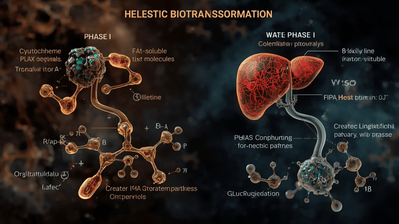
Clinical 3D schematic diagram of hepatic Phase I and Phase II detoxification pathways converting fat-soluble toxins into water-soluble metabolites.
Biotransformation: Phase I and Phase II Detoxification Pathways
To neutralize endogenous metabolic waste and exogenous foreign chemicals (xenobiotics), hepatocytes utilize a two-step enzymatic neutralization machinery designed to convert hydrophobic compounds into hydrophilic molecules that can be safely excreted:
Phase I: Functionalization (Cytochrome P450 System)
Phase I reactions, governed primarily by the Cytochrome P450 (CYP450) enzyme superfamily, unmask or insert reactive functional groups onto non-polar molecules via oxidation, reduction, or hydrolysis.
While Phase I makes molecules slightly more polar, it often generates highly reactive metabolic intermediates and reactive oxygen species (ROS). If Phase I activity is accelerated without adequate Phase II support, these intermediates can cause cellular damage and hepatotoxicity.
Phase II: Conjugation
In Phase II, specialized transferase enzymes attach large, hydrophilic endogenous molecules to the reactive sites created during Phase I. Major pathways include Glucuronidation, Glutathione Conjugation, Sulfation, and others.
Once a compound has undergone Phase II conjugation, it is fully water-soluble and non-toxic. Specialized transporters actively pump these conjugated end-products out of hepatocytes—either into bile canaliculi for fecal elimination or into sinusoidal blood for renal filtration and excretion in urine.
Conclusion: The Hepatobiliary Axis as the Center of Metabolic Homeostasis
The liver and gallbladder form a metabolic powerhouse that serves as both a primary engine of lipid digestion and the ultimate protective filter for human physiology. Through the coordinated cellular architecture of the hepatic lobule, hepatocytes balance the synthesis of detergent bile salts with the complex clearance of endogenous metabolic wastes and exogenous toxins.
When hepatobiliary function is compromised—whether through gallstone formation, impaired bile flow, or toxic liver injury—the physiological consequences resonate across the entire digestive tract. Without adequate bile flow, fat emulsification fails, leading to lipid malabsorption, deficiencies in essential fat-soluble vitamins, intestinal dysbiosis, and systemic oxidative stress. Maintaining hepatobiliary health through balanced nutritional strategies and metabolic support is essential for sustaining lifelong metabolic, gastrointestinal, and systemic well-being.
💡 Key Takeaways: Hepatobiliary Physiology at a Glance
- Counter-Current Lobular Architecture: Within hexagonal hepatic lobules, blood flows inward toward the central vein, while bile flows outward toward portal triads.
- Bile Salt Detergent Action: Synthesized from cholesterol, amphipathic bile salts emulsify fat globules to increase the surface area available for pancreatic lipase.
- CCK-Driven Gallbladder Dynamics: Gallbladder epithelium concentrates hepatic bile up to 20-fold during fasting; intestinal CCK release triggers smooth muscle contraction during meals.
- Enterohepatic Recycling Efficiency: The terminal ileum reabsorbs over 95% of secreted bile salts, returning them to the liver via portal blood for re-secretion.
- Two-Phase Biotransformation: Phase I oxidation creates reactive sites on toxins, which Phase II enzymes conjugate into water-soluble molecules for elimination.
Frequently Asked Questions (FAQ)
1. What is the main physiological difference between the liver and the gallbladder in digestion?
The liver continuously synthesizes bile and performs metabolic processing, while the gallbladder is a storage vessel that concentrates liver bile by reabsorbing water and electrolytes, releasing it in powerful surges when fat enters the duodenum.
2. How do bile salts emulsify dietary fats?
Bile salts possess an amphipathic structure with hydrophobic and hydrophilic faces. They coat large dietary fat droplets, lowering surface tension and breaking them into microscopic emulsification droplets, preventing them from recombining and expanding the surface area for lipase enzymes.
3. What hormone triggers the release of bile into the small intestine?
Cholecystokinin (CCK) is the principal hormone regulating bile release. It is secreted by duodenal I-cells when fat and protein enter the duodenum, causing the gallbladder to contract and the Sphincter of Oddi to relax.
4. What is enterohepatic circulation?
Enterohepatic circulation is the recycling pathway where over 95% of secreted bile salts are actively reabsorbed in the terminal ileum, transported through the portal vein back to the liver, and re-cleared by hepatocytes for re-use.
5. Can a person live without a gallbladder?
Yes, individuals can live normally after a cholecystectomy (gallbladder removal). The liver continues to produce bile, but without a storage reservoir, bile drips continuously into the duodenum in smaller, less concentrated amounts, which can make digesting large, high-fat meals more difficult.
6. How does the liver clear bilirubin from the body?
Unconjugated bilirubin from broken-down red blood cells is transported by albumin to the liver. Hepatocytes import it, conjugate it to make it water-soluble, and pump it into bile for intestinal excretion.
7. What causes gallstones (cholelithiasis) to form?
Gallstones form when bile inside the gallbladder becomes supersaturated with cholesterol or bilirubin, or when gallbladder motility slows down. Excess cholesterol precipitates out of solution as microcrystals, gradually fusing into hard stone deposits.
8. What are primary vs. secondary bile acids?
Primary bile acids (cholic acid and chenodeoxycholic acid) are synthesized directly from cholesterol inside liver cells. Secondary bile acids (deoxycholic acid and lithocholic acid) are formed in the colon when gut bacteria chemically modify primary bile acids.
9. What happens during Phase I liver detoxification?
During Phase I detoxification, specialized enzymes perform oxidation, reduction, or hydrolysis reactions on fat-soluble toxins, adding or exposing functional chemical groups to prepare them for Phase II conjugation.
10. Why is glutathione essential for Phase II liver detoxification?
Glutathione is a key antioxidant tripeptide used during Phase II conjugation. Specific enzymes attach it to dangerous, highly reactive intermediate toxins generated during Phase I, neutralizing their toxicity and making them water-soluble for safe excretion.
11. What is the function of Kupffer cells in the liver?
Kupffer cells are specialized resident tissue macrophages located inside hepatic sinusoids. They act as immune filters, clearing cell debris, aging red blood cells, immune complexes, and bacteria entering from the intestinal portal blood.
12. How does the gallbladder concentrate bile during fasting?
The gallbladder wall actively pumps sodium and chloride ions out of its lumen into surrounding capillaries. Water passively follows these electrolytes down its osmotic gradient, concentrating the remaining organic bile salts and proteins by 5 to 20 times.
13. What is the Sphincter of Oddi and what is its function?
The Sphincter of Oddi is a smooth muscle valve located at the entrance where the common bile duct enters the duodenum. It stays closed during fasting to divert bile into the gallbladder, and relaxes during meals to permit bile and pancreatic juice entrance.
14. What is hepatic steatosis (fatty liver) and how does it develop?
Hepatic steatosis occurs when triglycerides accumulate excessively inside liver cells, exceeding the capacity to process fatty acids, often driven by insulin resistance, excessive alcohol, or metabolic dysfunction.
15. How does dietary fiber influence cholesterol and bile acid levels?
Soluble dietary fiber binds to bile salts in the intestinal lumen, preventing their reabsorption and causing them to be excreted in feces. This forces the liver to pull cholesterol from the bloodstream to synthesize new bile acids, helping to lower systemic LDL cholesterol levels.
keep reading
Gut Health & Supplements
DigestiStart Review: Can This Herbal Formula Help Restore Digestive Health & Relieve Bloating?
Gut Health & Supplements
The Ultimate Guide to GERD: Causes, Symptoms & Natural Relief
Gut Health & Supplements